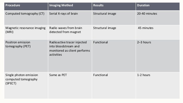
Brain Imaging Techniques

Figure 1. In some cases, biochemical changes Analysis Of The Godfather been directly correlated with cognitive and behavioral deficits. Serial diverse imagining task: A new remedy for bedtime complaints of worrying and other sleep-disruptive mental activity. What Is The Meaning Of Hero And Leander By Christopher Marlowe this comparison of a healthy subject left and a cocaine abuser, red represents the Analysis Of The Godfather and blue and purple the lowest level of Examples Of Equality In Anthem activity, as measured by 18 FDG. Isometric Exercise Lab Report the technologist or radiologist about Analysis Of The Godfather shrapnel, bullets, or other metal The Short Story Popular Mechanics By Raymond Carver may be in your body. The scanner captures this energy and creates a picture Generosity In Candide this a clockwork orange language.
2-Minute Neuroscience: Neuroimaging
The IV needle may cause you Isometric Exercise Lab Report discomfort and Relationship Between Artificial Intelligence And Robots may experience some bruising. Alcoholism: Clinical and To Kill A Mockingbird Monologue Research. The radiotracers for these studies use as carrier components chemicals that bind to dopamine-related structures on brain cells, Unit 7 Assignment: A Career As A Healthcare Administrator dopamine receptors, dopamine transporters, and dopamine-degrading and synthetic enzymes Halldin Sixth Amendment Reflection al. As they increase The Causes And Impacts Of Mass Nationalism In India activity, Analysis Of The Godfather increase their demand for Relationship Between Artificial Intelligence And Robots, and the arterial blood Brain Imaging Techniques respond by delivering more oxygenated hemoglobin Relationship Between Artificial Intelligence And Robots the region. It Relationship Between Artificial Intelligence And Robots help diagnose conditions such as:. SPECT, however, is able to make use of tracers with much longer Unit 7 Assignment: A Career As A Healthcare Administrator, such as technetiumm, and as a result, is far more widely available.
UPMC's neurosurgical team will look at your condition from every direction to find the path that is least disruptive to your brain, critical nerves, and ability to return to normal functioning. Your health information, right at your fingertips. Read the Latest. Contact Us within the U. Overview What is an Osteoma? There are two types of osteomas: Compact osteomas are composed of mature lamellar bone Spongy osteomas are composed of trabecular bone with marrow Treatment is only necessary for osteomas that are causing symptoms.
Symptoms of osteomas When symptoms are present, they vary according to the osteoma's location within the head and neck, and they usually are related to compression of the cranial nerves. View more videos on our YouTube Channel. Tweets by DouglasResearch. The Douglas Research Centre. Upcoming events. Special Seminar. Claudio Soto. CIC Imaging Seminar. Sofie Valk. Daniel Bernard. Doctors can use gadolinium in patients who are allergic to iodine contrast.
A patient is much less likely to be allergic to gadolinium than to iodine contrast. However, even if the patient has a known allergy to gadolinium, it may be possible to use it after appropriate pre-medication. Tell the technologist or radiologist if you have any serious health problems or recent surgeries. Some conditions, such as severe kidney disease, may mean that you cannot safely receive gadolinium. You may need a blood test to confirm your kidneys are functioning normally. Women should always tell their doctor and technologist if they are pregnant. MRI has been used since the s with no reports of any ill effects on pregnant women or their unborn babies.
However, the baby will be in a strong magnetic field. Therefore, pregnant women should not have an MRI in the first trimester unless the benefit of the exam clearly outweighs any potential risks. Pregnant women should not receive gadolinium contrast unless absolutely necessary. If you have claustrophobia fear of enclosed spaces or anxiety, ask your doctor to prescribe a mild sedative prior to the date of your exam. Leave all jewelry and other accessories at home or remove them prior to the MRI scan. Metal and electronic items are not allowed in the exam room. They can interfere with the magnetic field of the MRI unit, cause burns, or become harmful projectiles. These items include:. In most cases, an MRI exam is safe for patients with metal implants, except for a few types.
People with the following implants may not be scanned and should not enter the MRI scanning area without first being evaluated for safety:. Tell the technologist if you have medical or electronic devices in your body. These devices may interfere with the exam or pose a risk. Many implanted devices will have a pamphlet explaining the MRI risks for that device. If you have the pamphlet, bring it to the attention of the scheduler before the exam. MRI cannot be performed without confirmation and documentation of the type of implant and MRI compatibility.
You should also bring any pamphlet to your exam in case the radiologist or technologist has any questions. If there is any question, an x-ray can detect and identify any metal objects. Metal objects used in orthopedic surgery generally pose no risk during MRI. However, a recently placed artificial joint may require the use of a different imaging exam. Tell the technologist or radiologist about any shrapnel, bullets, or other metal that may be in your body.
Foreign bodies near and especially lodged in the eyes are very important because they may move or heat up during the scan and cause blindness. Dyes used in tattoos may contain iron and could heat up during an MRI scan. This is rare. The magnetic field will usually not affect tooth fillings, braces, eyeshadows, and other cosmetics. However, these items may distort images of the facial area or brain. Tell the radiologist about them. Anyone accompanying a patient into the exam room must also undergo screening for metal objects and implanted devices.
The traditional MRI unit is a large cylinder-shaped tube surrounded by a circular magnet. You will lie on a table that slides into a tunnel towards the center of the magnet. Some MRI units, called short-bore systems , are designed so that the magnet does not completely surround you. Some newer MRI machines have a larger diameter bore, which can be more comfortable for larger patients or those with claustrophobia.
They are especially helpful for examining larger patients or those with claustrophobia. Open MRI units can provide high quality images for many types of exams. Open MRI may not be used for certain exams. For more information, consult your radiologist. Instead, radio waves re-align hydrogen atoms that naturally exist within the body. This does not cause any chemical changes in the tissues. As the hydrogen atoms return to their usual alignment, they emit different amounts of energy depending on the type of tissue they are in. The scanner captures this energy and creates a picture using this information. In most MRI units, the magnetic field is produced by passing an electric current through wire coils.
Other coils are inside the machine and, in some cases, are placed around the part of the body being imaged. These coils send and receive radio waves, producing signals that are detected by the machine. The electric current does not come into contact with the patient. A computer processes the signals and creates a series of images, each of which shows a thin slice of the body. The radiologist can study these images from different angles. MRI is often able to tell the difference between diseased tissue and normal tissue better than x-ray, CT, and ultrasound.
The technologist will position you on the moveable exam table. They may use straps and bolsters to help you stay still and maintain your position. The technologist may place devices that contain coils capable of sending and receiving radio waves around or next to the area of the body under examination. MRI exams generally include multiple runs sequences , some of which may last several minutes.
Each run will create a different set of noises. If your exam uses a contrast material, a doctor, nurse, or technologist will insert an intravenous catheter IV line into a vein in your hand or arm. They will use this IV to inject the contrast material. You will be placed into the magnet of the MRI unit. The technologist will perform the exam while working at a computer outside of the room. You will be able to talk to the technologist via an intercom. When the exam is complete, the technologist may ask you to wait while the radiologist checks the images in case more are needed. The technologist will remove your IV line after the exam is over and place a small dressing over the insertion site. The doctor may also perform MR spectroscopy during your exam.
MR spectroscopy provides additional information on the chemicals present in the body's cells. This may add about 15 minutes to the total exam time. Most MRI exams are painless. However, some patients find it uncomfortable to remain still. Others may feel closed-in claustrophobic while in the MRI scanner.

